Introduction to Imaging Mass Spectrometry
One-day course

Instructor:
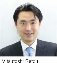
Mitsutoshi Setou, Hamamatsu University School of Medicine, Hamamatsu, Shizuoka, Japan
Schedule:
Sat., Sep. 15, 2012, 10.00 am - 17.00 pm
Matrix-assisted laser desorption/ionization (MALDI)-imaging mass spectrometry (IMS) is emerging as a powerful tool for investigating the distribution of molecules, such as proteins and metabolites, within biological systems through the direct analysis of thin tissue sections. Unique among imaging methods, MALDI-IMS can determine the distribution of hundreds of unknown compounds in a single measurement and identify the molecules on tissue sections. Recently, Mass Microscopes were introduced to the research community, enabling us to detect conventional molecules in tissue structures by optical microscopes with an objective lens, as well as volatile ones using an atmospheric ion source chamber. Altogether, this offers the opportunity to visualize molecular distributions with more high-spatial resolution by the minimum laser diameter (< 10 m) and identify them from their individual fragmentation patterns. Moreover, our group together with the Japanese foundation for cancer research have developed an analyzing software for IMS datasets. Using this software, we can easily extract meaningful signals based on common and/or specific expressions from enormous IMS datasets. This course covers the fundamental principal and techniques of MALDI-IMS, assuming no previous experience in IMS or basic mass spectrometry. The following topics are covered: introduction to principles and applications of IMS technology, such as making tissue sections, applying matrices onto tissue sections, acquiring IMS datasets from tissue sections, creating ion images from the IMS dataset, and statistical analyses of data.
Maximum capacity of participants:
50
Fee:
Regular 10,000 JPY; Student 5,000 JPY.


